

Diagram Of A Plant Cell
Definition of constitute cell
Found cells are eukaryotic cells, that are found in greenish plants, photosynthetic eukaryotes of the kingdom Plantae which means they accept a membrane-bound nucleus. They have a variety of membrane-jump prison cell organelles that perform various specific functions to maintain the normal operation of the constitute cell.
Construction of Plant prison cell
Generally, plant cells are a lot bigger than animal cells, coming in more like sizes and they are typically cubed or rectangular in shape. Plant cells also have structural organelles that are non institute in the animals' cells including the jail cell wall, vacuoles, plastids e. one thousand Chloroplast. Animal cells likewise contain structures that are non establish in the plant cells such every bit, cilia and flagella, lysosomes, and centrioles.
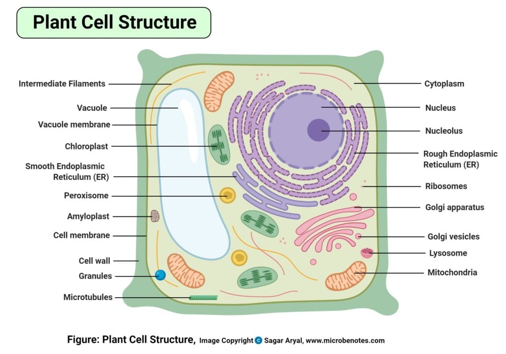
Figure: Labeled diagram of plant cell, created with biorender.com
The typical characteristics that define the plant cell include cellulose, hemicellulose and pectin, plastids which play a major role in photosynthesis and storage of starch, big vacuoles responsible for regulating the prison cell turgor pressure. They also have a very unique prison cell division process whereby there is the formation of a phragmoplast (a complex made up of microtubules, microfilaments, and the endoplasmic reticulum) all assembling during cytokinesis, to divide the daughter cells.
These organelles most of them are similar to the creature organelles performing the same functions equally those of the animal cell. Organelles accept a wide range of responsibilities that include everything from producing hormones and enzymes to providing energy for a establish cell.
Plants cells have Dna that helps in making new cells, hence enhancing the growth of the found. the DNA is enclosed within the nucleus, an enveloped membrane structure at the center of the cell. The plant cell also has several prison cell organelle structures performing a multifariousness of functions to maintain cellular metabolisms, growth, and development.
Plant Jail cell Gratuitous Worksheet
Answer primal
List of Plant jail cell organelles
- Prison cell Wall
- Cytoskeleton
- Cell (Plasma) membrane
- Plasmodesmata
- The cytoplasm
- Plastids
- Constitute Vacuoles
- Mitochondria
- Endoplasmic reticulum (ER)
- Ribosomes
- Storage granules
- Golgi bodies
- Nucleus
- Peroxisomes
Plant Prison cell Wall
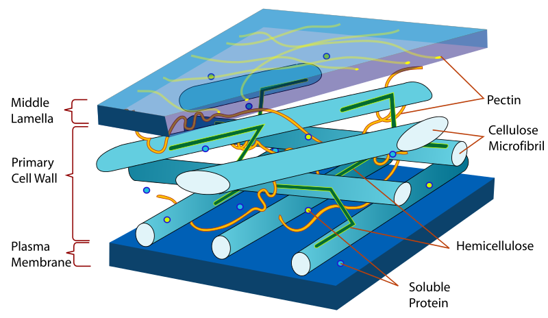
Figure: Diagram of Plant prison cell wall. Source: Wikipedia
Definition of plant cell wall
It is the rigid outer cover of the institute jail cell with a major part of protecting the plant cell, giving it, its shape.
Construction of plant cell wall
- Information technology is a specialized matrix that covers the surface of the plant cell. Every constitute cell has a cell wall layer which is a major distinguishing factor between a plant prison cell and an animal cell.
- The jail cell wall is fabricated upwards of two layers, a middle lamella, and a primary cell wall and sometimes a secondary cell wall.
- The eye lamella acts as the strengthening layer between the primary walls of the neighboring cells.
- The main wall is made up of cellulose underlying the cells that are dividing and maturing. The chief wall is a lot thinner and less rigid every bit compared to those of the cells that accept reached complete maturation. The thinness allows the cell wall to aggrandize.
- After full cell growth, some plants get rid of the primary wall but almost, they thicken the principal wall or it makes some other layer with rigidity but a different arrangement, known equally the secondary wall.
- The secondary wall offers permanent stiff mechanical support to the plant prison cell peculiarly the support found in wood.
- In contrast to the permanent stiffness and load-bearing capacity of thick secondary walls.
The function of the establish jail cell wall
The chief role of the jail cell wall is divers to exist a mechanical and structural function, that is highly constructive in serving the plant cell. These functions include:
- Providing the cell with mechanical protection and shielding the prison cell from the chemically harsh surround, provided by the secondary wall layer.
- It is semipermeable hence it allows in and out, the apportionment of materials such as water, molecular nutrients, and minerals.
- It too forms provides a rigid building block to stabilize the plant to produce some of its structures, for example, the stem and leaves of the plants.
- Information technology also provided a site for the storage of some elements such as the regulatory molecules that detect pathogens in the constitute, hindering the evolution of diseased tissue.
- The thin primary walls serve as structural and supportive functional layers when the cell vacuoles are filled with water, exerting turgor pressure on the cell wall, thus maintaining the plants' stiffness and preventing plants from losing water and withering.
The basic edifice block is made of cellulose fibers, of both the primary and secondary walls, despite having different compositions and structures. Cellulose is a polysaccharide matrix that offers tensile strength to the cells. This forcefulness is entrenched within the highly full-bodied matrix of water and glycoproteins.
Found cytoskeleton
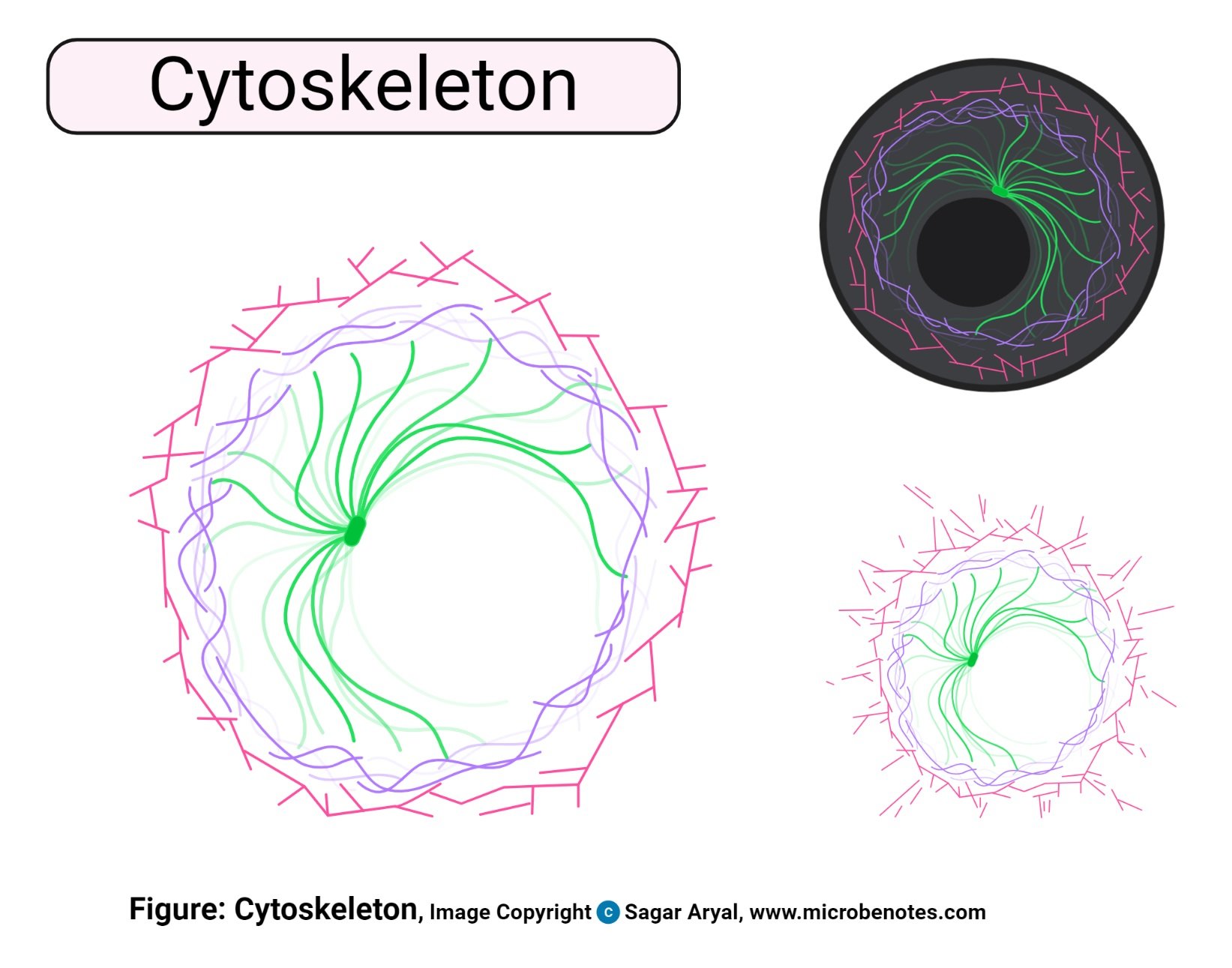
Effigy created with biorender.com
Definition of the constitute cytoskeleton
This is a network of microtubules and filaments that plays a primary role in maintaining the plant cell shape and giving the cell cytoplasm support and maintaining its structural organization. These filaments and tubules commonly extend all over the cell, through the jail cell cytoplasm. Besides giving support and maintaining the jail cell and the cell cytoplasm, its as well involved in the transportation of cellular molecules, cell division, and jail cell signaling activities.
Construction of the establish cytoskeleton
The cytoskeleton has an essential definition of the structure of eukaryotic cells, describing the back up system of these cells, the maintenance factors and transport involvements within the prison cell. These functions are defined by the structure of the cytoskeleton which is fabricated upwardly of iii filaments i. e actin filament (microfilaments), microtubules and intermediate filaments.
- Microfilaments, besides known as actin filaments, are a meshwork of fibers running parallel to each other. They are made up of the thin strands of actin proteins hence the name actin filaments. They are the thinnest filaments of the cytoskeleton with a thickness of 7 nanometers.
- Intermediate filaments have a diameter of almost viii-12 nm; They lie between the actin filaments and the microtubules. Its function in plant cells is not clearly understood
- Microtubules are hollow tubes made up of tubulins, with a bore of 23nm. They are the largest filament compared to the other two filaments.
Functions of the plant cytoskeleton
Microfilaments
- They play a main function is a sectionalisation of the cell cytoplasm past a mechanism known every bit cytokinesis, forming 2 girl cells.
- They also participate in cytoplasmic streaming, a procedure of cytosol menstruation all over the cell, transporting nutrients and cell organelles.
Intermediate Filaments
- The intermediate filaments' function in the plant cells is not clearly understood but has a role to play in maintaining the cell shape, structural support and retain tension within the jail cell.
Microtubules
- Unlike the part of the microtubule in cell division in the animal cell, the plant cell uses the microtubules to transport materials within the vell and they are also used in forming the plant prison cell, cell wall.
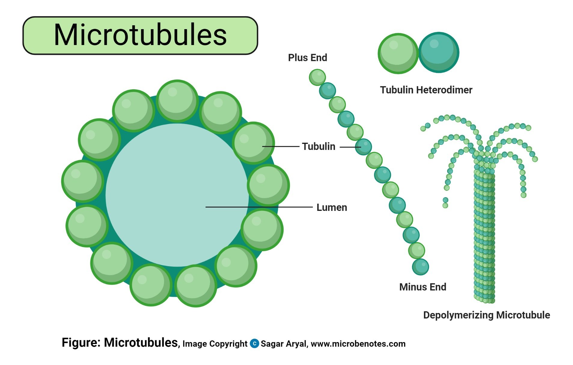
Figure created with biorender.com
Other functions of the cytoskeleton in plants include:
- Giving the plant cell shape, maintaining the cell shape and transportation of some cell organelles throughout the prison cell, molecules, and nutrients across the cell cytoplasm.
- Information technology also plays a function in mitotic cell sectionalisation.
- In summary, the cytoskeleton is the frame of building the cell, hence it maintains the cell structure, provides cell structural back up and defines the prison cell structure.
Chloroplast of constitute cell
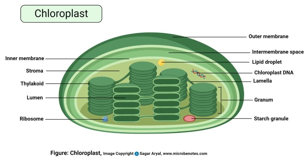
Effigy: Diagram of chloroplast, created with biorender.com
Structure of the plant jail cell chloroplast
- These are organelles establish in found cells and algal cells.
- They are oval-shaped.
- They are fabricated up of two surface membranes, i.e outer and inner membrane and an inner layer known as the thylakoid layer has 2 membranes.
- The outer membrane forms the external lining of the chloroplast while the inner membrane is below the outer layer.
- The membranes are separated past thin membranous space and within the membrane, in that location is also a space known as the stroma. The stroma houses the chloroplast.
- The third layer known as the thylakoid layer is extensively folded making the appearance of a flattened disk known as thylakoids which have big numbers of chlorophyll and carotenoids and the electron transport concatenation, defined as the 50ight-harvesting complex, used during photosynthesis.
- Thylakoids are piled on top of each other in stacks known equally grana.
Functions of the plant prison cell chloroplast
- The chloroplast is the site of food synthesis for establish cells, by a mechanism known as photosynthesis.
- Chloroplasts comprise chlorophyll, a green pigment that absorbs calorie-free energy from the lord's day for photosynthesis.
- The photosynthesis procedure converts water, carbon dioxide, and lite energy into nutrients for utilization by the plants.
- Thylakoids contain chlorophyll pigments and carotenoids for trapping light free energy for use in photosynthesis.
- the chlorophyll pigment gives plants their green color.
Chromoplast plastid of the plant cell
Chromoplast definition
- Chromoplasts ascertain all the found pigments stored and synthesized in plants. They are found in a variety of plants of all kinds of ages.
- They are normally formed from the chloroplasts is the name given to an area for all the pigments to be kept and synthesized in the plant.
- The accept carotenoid pigments that allow the differentiation in color seen in flowers and fruits. Its color attracts pollination mechanisms by pollinators.
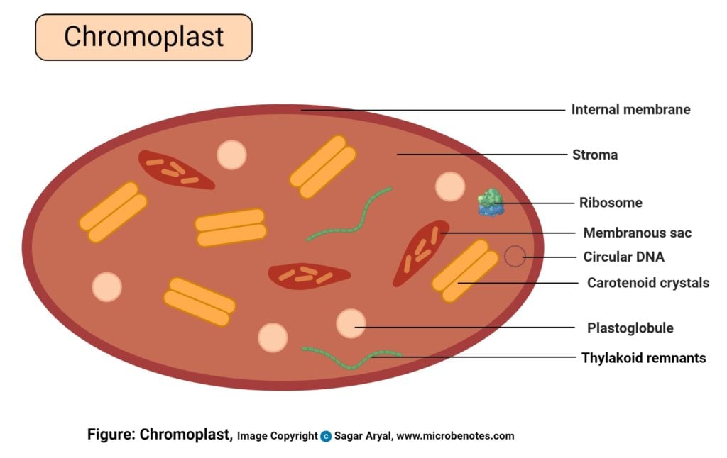
Effigy: Diagram of chromoplast, created with biorender.com
Structure of plant chromoplast
Microscopic ascertainment indicates that chromoplast has at least four types:
- Proteic stroma which contains granules
- Amorphous paint with granules
- Protein and pigment crystals
- Crystalised chromoplast
Although, the more specialized feature has been observed classifying information technology further into 5 types:
- Globular chromoplasts which appear every bit globules
- Crystalline chromoplast which appears crystalized
- Fibrillar chromoplast which appears like fibers
- Tubular chromoplast which looks similar tubes
- Membranous chromoplast
These chromoplasts live amid each other though some plants have specific types such every bit mangoes accept the globular chromoplast while carrots have crystallized chromoplast, tomatoes have both crystalline and bleary chromoplast because they accumulate carotenoids.
Functions of constitute chromoplast
- They give distinctive colors to institute parts such as flowers, fruits, roots, and leaves. Differentiation of chloroplast to chromoplast makes the fruits of plant ripen.
- They synthesize and store plant pigments such every bit yellowish pigments for xanthophylls, orange for carotenes. This gives the found and its parts the color.
- They attract pollinators by the colors they produce, which helps in the reproduction of the plant seed.
- Chromoplats found in roots enable the aggregating of water-insoluble elements specially in tubers such as carrots and potatoes.
- They contribute to color alter during found aging, for flowers, fruits, and leaves.
Gerontoplast plastids of the constitute cell
- These plastids found in plant leaves are the organelles responsible for cell aging. They differentiate from chloroplast when the plants start to age, and they tin can not perform photosynthesis anymore.
- They appear as unstacked chloroplasts without a thylakoid membrane and aggregating of plastoglobuli that is used in producing energy for the cell.
- The main function of Gerontoplast is to aid the aging of the plant parts giving them a distinct color to indicate a lack of photosynthesis procedure.
Leucoplast plastids of the plant cell
- These are the not-pigmented plastids. Since they lack the chloroplast pigments, they are found in not-photosynthetic parts of the plants like the roots and seeds.
- They are smaller than the chloroplasts, which varying morphologies others appearing ameboid shaped.
- They are interconnected with a network of stromules in roots, flower petals.
- They can be specialized to store starch, lipids, and proteins in big quantities hence named as amyloplasts, elaioplast, and proteinoplast, depending on what they store respectively.
The primary role of the leucoplast includes:
- Storage of starch, lipids, and proteins.
- They are also used to convert amino acids and fatty acids.
Plant Vacuoles
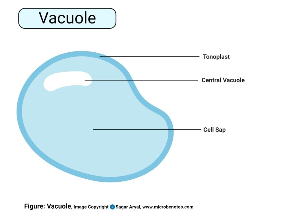
Figure created with biorender.com
Constitute vacuoles definition
- Plant cells accept large vacuoles as compared to animal cells.
- The central vacuoles are found in the cytoplasmic layer of cells of a diverseness of different organisms, but larger in the establish cells.
Structure of establish prison cell vacuoles
- These are big, vesicles filled with fluid, inside the cytoplasm of a cell.
- It is made upwardly of thirty% fluid of the cell volume merely can fill upwards to ninety% of the cell'south intracellular space.
Functions of the central vacuole
- The fundamental vacuoles are used to adjusted the size of the cell and to maintains the turgor pressure level of the plant cells, preventing wilting and withering of plants particularly the leaves.
- When the cytoplasmic book is constant, the vacuoles account majorly for the size of the institute cell.
- Turgor pressure is maintained when the vacuoles are full of water. When there is no turgor pressure level, information technology is an indication of the establish losing water, hence the plant leaves and stems wither.
- Plant cells thrive in high water levels (Hypotonic solutions), taking up water by osmosis from the environment, thus maintaining turgidity.
- A constitute cell can have more one type of vacuole. some specialized vacuoles especially those structurally related to lysosomes contain degradative enzymes used to break down macromolecules.
- Vacuoles are also responsible for the storage of cellular nutrients including sugars, organic salts, inorganic salts, proteins, cellular pigments, lipids. these elements are stored until when the cell requires them for cellular metabolisms. For example, vacuoles store proteins for seeds and opium metabolites.
Mitochondria of the found jail cell
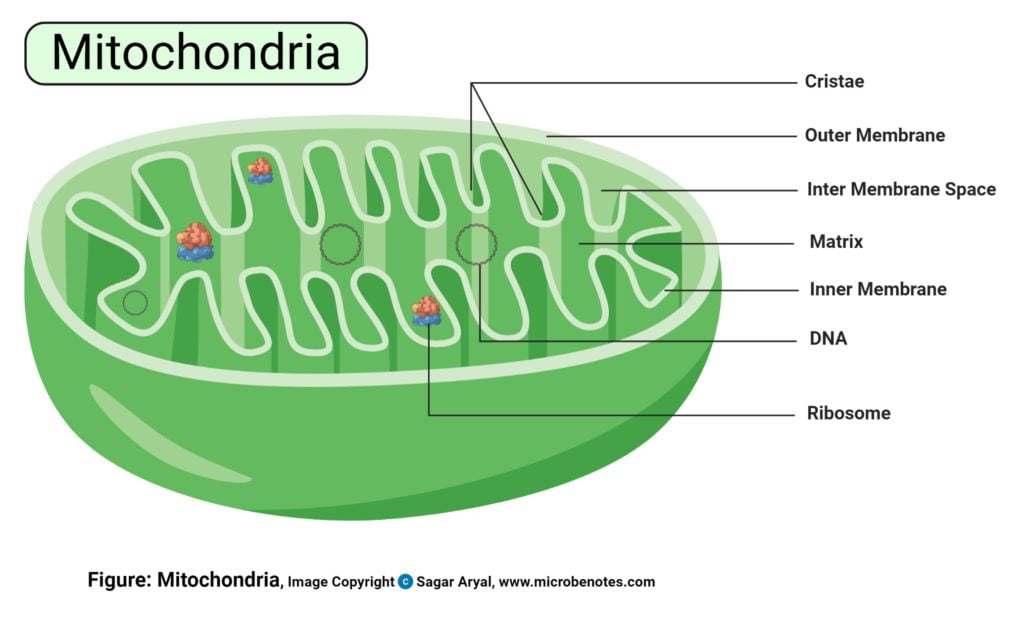
Figure created with biorender.com
Establish prison cell mitochondria definition
- Mitochondria are too known every bit chondriosomes, are the ability generating organelles of a jail cell, hence they are commonly known as the powerhouse of the cell.
- The mitochondria convert stored nutrients by the assistance of oxygen to produce free energy in for of (ATP )Adenosine TriPhosphate, hence they are the site for not-photosynthetic energy transduction.
- There are hundreds of mitochondria within a single plant jail cell.
- Mitochondria are plant in high numbers within the phloem paint of the institute jail cell, and the neighboring cells have high metabolism rates. This is to supply energies that support various needing mechanisms, similar the transportation of food through the sieve tubes.
- As they perform their mechanisms, mitochondria continuously motility and alter their shapes, depending on its interactions with light trapped for photosynthesis, level of cytosolic sugars and the endoplasmic reticulum mediated interactions.
- The animal and plant mitochondria are very similar except for a few notable differences east.1000. mitochondria in plants accept reduced nicotinamide adenine dinucleotide (NADH) dehyg=drogenase used for oxidation of exogenous NADH which animal cell lack.
- Mitochondria from many establish sources are relatively insensitive to cyanide inhibition, a feature not found in creature mitochondria. On the other hand, the b -oxidation pathway of fatty acids is located in fauna mitochondria, whereas in plants, the enzymes of fatty acid oxidation occur in the glyoxysomes. (https://publishing.cdlib.org/ucpressebooks/view?docId=ft796nb4n2&chunk.id=d0e6787&toc.depth=1&toc.id=d0e6787&make=ucpress)
Structure of plant mitochondria
- Plant cell mitochondria have loftier pleomorphism.
- Mitochondria in green plants are discrete, spherical-oval shaped organelles of diameter ranging from 0.2to1.5μm
- The mitochondria take a double-layered system i. e a polish outer membrane and an inner complex membrane that encloses the organelle matrix.
- The two layers are lipid bilayers complexed with a hydrophobic fatty acid chain. These lipids are a form of phospholipids that are highly dynamic with a strong attraction to the fatty acrid regions.
- They have a mitochondrial gel-matrix in the central mass.
- The mitochondria also possess all the enzymes for the Tricarboxylic cycle (TCA) including citrate synthetase, Pyruvate oxidase, Isocitrate Dehydrogenase, Malate Dehydrogenase, Malic Enzyme.
Functions of mitochondria in plants
- The mitochondria are the powerhouse of the cell, hence their major office is generating free energy for use by the jail cell.
- To have a high rate of metabolism because they supply energy for the unknown machinery past which foods, mainly sucrose, are transported in the sieve tubes.
- Within the mitochondria, the potential energy in food that is manufactured by photosynthesis is what is used for the metabolisms of the cells. For example, energy used for the formation of new cell content, enzyme production and moving of sugar molecules are produced by the mitochondria.
- This is the cite for the Tricarboxylic cycle (TCA), also known equally the Krebs wheel. The TCA wheel uses the jail cell's nutrients, converting them into past-products that the mitochondria use for producing energy. These processes accept place in the inner membrane because the membrane bends into folds called the cristae, where the poly peptide components used for the main free energy product system cells, known as the Electron Transport Concatenation (ETC). ETC is the main source of ATP production in the body.
Endoplasmic reticulum (ER) of the establish cell
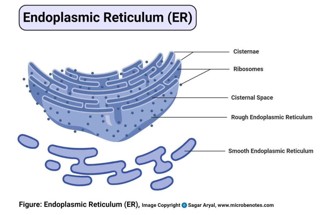
Effigy created with biorender.com
Found cell endoplasmic reticulum (ER) definition
- The ER is a continuous network of folded membranous sacs housed in the jail cell cytosol. It is a complex organelle taking upward a sizable part of the jail cell'south cytosol
- It is made upwardly of two regions known as the rough endoplasmic reticulum (they take ribosomes attached to their surface membrane) and the smooth endoplasmic reticulum (they lack ribosomal attachment).
- The endoplasmic reticulum known for its high dynamics functions in eukaryotic cells, play major roles in synthesizing, processing, transporting and storing proteins, lipids, and chemic elements. These elements are used by the constitute cell and other organelles such as the vacuoles and the apoplast (Plasma membrane).
- The inner infinite of the ER is known as the lumen.
- It is fastened to the nuclear envelope, providing a link betwixt the nucleus and the jail cell cytosol, and also giving a link between the cell to the plasmodesmata tubes, which connect to the plant cells. It accounts for 10% of the volume of the cytosol.
- On the other mitt, rough ER virtually e'er appears equally stacks of double membranes that are heavily dotted with ribosomes. Based on the consistent appearance of crude ER, it most likely consists of parallel sheets of membrane, rather than the tubular sheets that narrate smooth ER.
- These flattened, interconnected sacs are called cisternae, or cisternal cells. The cisternal cells of rough ER are also referred to as luminal cells. Rough ER and the Golgi complex are both composed of cisternal cells.
Structure of found cell endoplasmic reticulum
- This is a consistently folded bleary organelle found in the cytoplasm of the jail cell, that is made up of a sparse network of flattened interconnected compartments (sacs) that connects from the cytoplasm to the prison cell nucleus.
- Inside its membranes, at that place are membranous spaces called thecristae spaces and the membrane folding are chosencristae.
- There are two types of ER based on their construction and the function they perform includingRough Endoplasmic reticulum and theShine endoplasmic reticulum.
Functions of the endoplasmic reticulum
Functions of the Crude and polish endoplasmic reticulum
- The Crude endoplasmic reticulum is covered by ribosomes around its surface membrane, making a rough bumpy appearance. the main role of the Rough ER in synthesizing proteins, which are transported from the prison cell to the Golgi bodies, which carry them to other parts of the plant to help in its growth. These proteins are an associates of amino acid sequences that combine to class antibodies, hormones, digestive enzymes. the assembling is accomplished by the ribosomes fastened to the rough ER.
- Some proteins are processed exterior the cell, they can also be transported into the Crude ER where they undergo assembling into the right shape and dimensions for cell utilization and conjugated with carbohydrate elements to class a complete poly peptide. these complexes are then transported and distributed to parts of the ER known every bit the transitional ER, for packaging in cell vesicles and passed to the Golgi bodies which export them to other parts of the plant.
- The shine ER is polish due to a lack of attached surface ribosomes. They look every bit though they are budding off from the lumen of the crude endoplasmic reticulum. Its part is synthesizing, secreting and storing lipids, metabolizing carbohydrates and manufacturing of new membranes. This is enhanced past the presence of several enzymes bound to its surface.
- When a found has enough free energy for utilization for photosynthesis and however possess excess lipids manufactured past the prison cell, these lipids are stored in the smooth Endoplasmic reticulum in the form of triglycerides. And when the cell needs more free energy, the triglycerides are broken down to produce the free energy required by the plants.
- Minimally, the smooth endoplasmic reticulum has also been linked to the formation of the cellulose on the prison cell wall.
Other functions of the endoplasmic reticulum in the plant cell
- Calcium is used in the growth and evolution of plant cells which enhances plant growth but in some cases, calcium may be produced in excessive quantities that harm the plant cell by causing cell decease. Therefore the Endoplasmic reticulum has been linked to regulating the excess calcium by converting information technology to calcium oxalate crystals. Specialized cells in the endoplasmic reticulum known as crystal idioblast play a major role in this conversion and as well in storing these crystals.
- The ER as well act as plant sensors. Plants have the ability to make rapid movements in response to sure external stimuli e. m low-cal intensity, temperature, and atmospheric pressure. In such mechanisms, the ER mediates for the plant to respond accordingly. For example, in Venus flytrap institute, react sensitively to touch, this is due to the presence of the cortical endoplasmic reticulum (Cortex cells) that instantly respond to bear upon.
- In the outcome of sensitivity, the sensory ER movement and collect at the top and the bottom of the cell, making them exist squeezed together thus causing a constraint on them. This leads to the release of accumulated calcium, which in turn produces the sense of touch.
- The cortical ER is highly linked with the plasmodesmata (a narrow thread of cytoplasm that passes through the cell walls of next institute cells and allows advice between them). The Plasmodesmata acts as a channel of communication among the cells thus linking to the motor cells triggering the cells and the establish to reply accordingly.
Ribosomes of the Found Cell
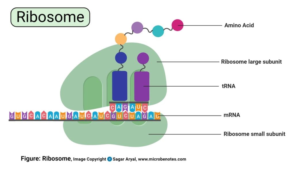
Figure created with biorender.com
Establish cell ribosome definition
- This is the organelle responsible for protein synthesis of the prison cell.
- Its found in the cell cytoplasm in large numbers and a few of them called functional ribosomes can be plant in the nucleus, mitochondria, and the cell chloroplast.
- Its made upwards of ribosomal DNA (rDNA) and cell proteins
- The procedure of poly peptide synthesis past the ribosomes is known as translation, by using the messenger RNA, which delivers the nucleotides to the ribosomes.
- The ribosomes then guide and interpret the message in the form of nucleotides, independent by the mRNA.
Structure of ribosomes of the establish cell
- The ribosomes' structure is the aforementioned in all cells merely smaller in prokaryotic cells. Generally, ribosomes in eukaryotic cells are big and they can only exist measured in Svedberg units (S). South unit of measurement is a measure of aggregation of large molecules to sediments on centrifugation. High S value means fast sedimentation charge per unit hence greater mass.
- Eukaryotic cell sediment in the 90s while prokaryotic cell sediment in the 70s.
- Ribosomes plant in the mitochondria and chloroplasts are equally small as the prokaryotic ribosomes.
- Naturally, ribosomes are made upward of two subunits i. e pocket-sized and large subunits, both classified according to their sedimentation rates past the Due south unit of measurement.
- The constitute prison cell, beingness a eukaryotic cell, has large complex ribosomes with higher S units, with four rRNAs with over fourscore proteins. The big subunit has the S unit of the 60s (28s rRNA, v.8s rRNA, and 5s rRNA) with 42 proteins. The pocket-sized subunit has a sedimentation rate of the 40s, made upwards one rRNA and 33 proteins.
- The ribosomal subunits combine in the nucleolus of the cell, which is and so transported into the cytoplasm through the nuclear pores. The cytoplasm is the master site for protein synthesis (translation).
Functions of ribosomes in found cells
- Containing a subunit of RNA, ribosomes major functions is to synthesize proteins for the cellular functions such as cell repair machinery.
- Ribosomes deed as catalysts in producing strong binding for portion extension using peptidyl transfer and peptidyl hydrolysis.
- Ribosomes institute in the jail cell cytoplasm are responsible for the conversion of genetic codes to amino acid sequences and edifice protein polymers from amino acid monomers.
- they are as well used in protein assembling and folding.
Storage granules of plant cell
- These are aggregates found within the cytoplasmic membrane and the plant cell plastids.
- They are inert organelles found in plants whose primary function is to store starch.
Functions of storage granules in institute jail cell
- They are used every bit food reservoirs
- They shop carbohydrates for the cell in the grade of glycogen or carbohydrate polymers
- They naturally shop starch granules for the plant cell
- They also fuel metabolisms in the prison cell that involved chemical reactions thus producing free energy for the product of new cellular materials.
Golgi bodies of constitute jail cell
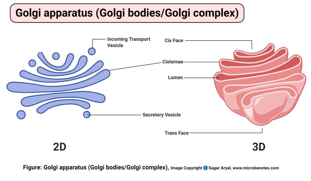
Figure created with biorender.com
Found jail cell Golgi bodies definition
- These are circuitous membrane-bound jail cell organelles found in the cytoplasm of a eukaryotic prison cell, which is also known every bit the Golgi complex or Golgi apparatus. They lie but next to the endoplasmic reticulum and near the nucleus.
Structure of the Golgi bodies in a establish cell
- Golgi bodies are maintained together by cytoplasmic microtubules and clasped by a protein matrix
- They are made upwards of flattened stacked pouches known as cisternae.
- Plant cells have a few hundreds of the Golgi bodies moving along the cell'due south cytoskeleton, over the endoplasmic reticulum equally compared to the very few plant in animal cells (1-2).
- The Golgi bodies have three primary compartments:
- Cis Golgi network is also known equally Goods inwards, are the cisternae the is closest to the endoplasmic reticulum. Too chosen the cis Golgi reticulum it is the entry area to the Golgi apparatus.
- The medial or the Golgi stack- this is the Main processing expanse, placed at the central layer of the cisternae
- Trans Golgi network is too known as the Goods outwards cisternae. This is the farthest cisternae endoplasmic reticulum from the endoplasmic reticulum.
Functions of the Golgi bodies in a plant jail cell
- The Golgi bodies have several functions linked to them, from being an adjacent organelle to the endoplasmic reticulum to where they evangelize the jail cell products to. They are found in the center of the cells' secretory pathway, as a membranous circuitous that primarily functions to process, distribute and store proteins for utilize past the found during stress responses and others in leguminous plants such every bit cereals and grains.
- The presence of the membranous sac compartments, perform various chemically related functions. As new proteins are transported out of the endoplasmic reticulum through the Golgi bodies, they pass through the three compartments each compartment producing a unlike reaction to the molecules, modifying them in diverse ways i.e.
- Cleaving the protein molecules to oligosaccharides bondage
- Attaching of carbohydrate moieties of different side chains to the protein elements
- Add-on of fatty acids and phosphate groups to the elements and removal of monosaccharides.
- The cell vesicles carrying poly peptide molecules from the endoplasmic reticulum into the cis compartment, where the product is modified, and so packaged into other vesicles which then transports it to the next compartment. The transportation is enhanced by marking the vesicle with a tag similar a phosphate grouping or special poly peptide molecules, leading information technology to its side by side endpoint.
- Finally, when the vesicles have transported the proteins and lipid molecules, the Golgi bodies are responsible for assembling the production and transporting it to the final destination. This is enhanced by the presence of enzymes in the plants' Golgi bodies, which attache to the sugar moieties to the proteins, packing them and transporting them to the cell wall.
Nucleus of Plant Jail cell
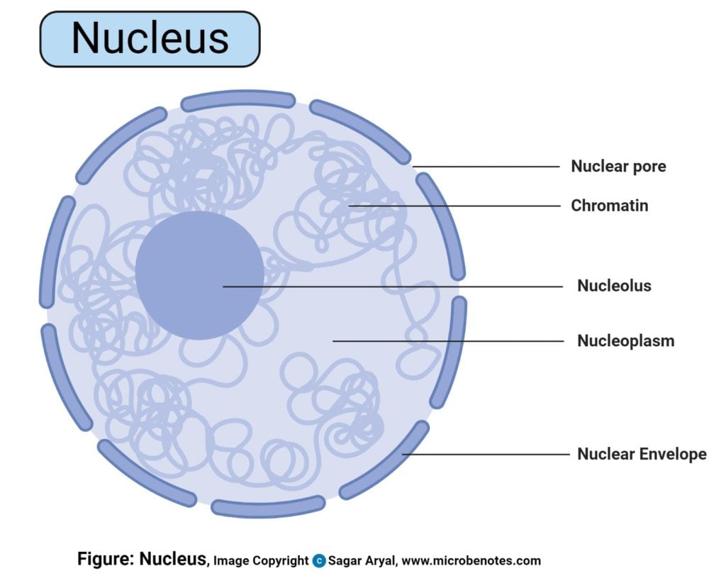
Figure created with biorender.com
Establish cell nucleus definition
- The nucleus is the information center of a jail cell. Information technology is a specialized complex organelle whose primary part is to store the cell'south genetic information.
- It is also responsible for coordinating the cell'south activities including cell metabolism, prison cell growth, synthesis of proteins and lipids and generally the cell reproduction by cell division mechanisms.
- The nucleus contains the cells' genetic data known equally Deoxyribonucleic acid (Dna), on the Chromosomes (special thread-like strands of nucleic acids and poly peptide institute in the nucleus, carrying genetic information)
Structure of the nucleus of the institute cell
- The nucleus is spherically shaped, centrally placed in the prison cell. It occupies about 10% of the prison cell book content.
- It as a double-layered membrane known every bit the nuclear envelope which separates the contents in the nucleus from those in the cell cytoplasm.
- The nuclear materials included chromatins, DNA which forms the cell chromosomes during jail cell division, the nucleolus which is responsible for synthesizing the cell ribosomes.
Functions of the nucleus of the plant cell
- The Master role of the cell nucleus is, it functions as the jail cell'south command center.
- The presence of the nuclear membrane, it encloses the nucleus and its contents from the cytoplasmic organelles. This nuclear membrane has the nuclear envelope, which has several nuclear pores, which offers selective permeability to and from the nucleus and the cytoplasm.
- The nucleus is also linked to the site for protein synthesis, i.due east the endoplasmic reticulum by a network of microfilaments and microtubules. These tubules extend all over the prison cell manufacturing elements and molecules depending on the specificity of the cell.
- Chromosomes: they are as well known as the chromatids. They are constitute in the cell nucleus of nigh all cells. They have vi long strands of Deoxyribonucleic acid which carve up into 46 separate molecules which pair upwardly into ii, fabricated of 23 molecules per chromosome. To grade a functional DNA unit, information technology is combined with cel proteins to form a compact structure of dense fiber-like strands known as the chromatins.
- The 6 DNA strands, each wraps around pocket-sized protein molecules produced past the ER known equally Histones. These class the beadlike structures known equally nucleosomes. Dna strands take a negative charge which is neutralized by the histones' positive charge. Unused DNA is folded and stored for future use.
Chromatins are classified into two types:
- Euchromatin: It is the active part of the Deoxyribonucleic acid that is used for RNA transcription producing cellular protein for cell growth and functioning.
- Heterochromatin: information technology is the inactive function of Dna that has the compressed and condensed DNA that is not in use.
During Chromatin germination, the chromatins change into other forms of the nucleus during cell division. Throughout the life of a cell, chromatin fibers take on unlike forms inside the nucleus. During the interphase stage of cell division, the euchromatin is expressed to get-go transcription. Into the metaphase stage, the chromatins divide making its own copies during replication exposing the chromatins more to form more than specialized structures known as chromosomes. These chromosomes so divide and split, forming ii new complete cells, with their ain genetic information.
Nucleolus
- It is a sub-organelle in the cell nucleus, which lacks a membrane.
- Its primary function is to synthesize the cell ribosomes, the organelles used to produce cellular proteins.
- The jail cell has about 4 nucleoli.
- The nucleolus is formed when chromosomes are brought together, merely before prison cell segmentation is initiated.
- The nucleolus disappears from during jail cell sectionalisation.
- The nucleolus is linked to cell crumbling which affects the aging of living things.
Nuclear Envelope
- Its made upward of two membranes separated from each other past perinuclear infinite. the infinite links into the endoplasmic reticulum.
- With its perforated wall, it regulates the molecules that enter and go out the nucleus into and out of the cytoplasm respectively.
- The inner membrane has a lining of proteins known as nuclear lamina, binding chromatins, and other nuclear elements.
- The envelope disintegrates and disappears during cell division.
Nuclear Pores
- They are perforate the prison cell envelope and their function is to regulate the passage of cellular molecules such equally proteins, histones through into and out of the nucleus and the cytoplasm respectively.
- They too allow Dna and RNA into the nucleus, providing free energy for making upward the genetic materials.
Peroxisomes of the Plant cell
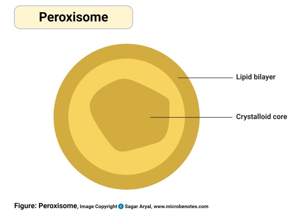
Figure created with biorender.com
Plant jail cell peroxisomes definition
These are highly dynamic tiny structures that take a single membrane containing enzymes responsible for the production of hydrogen peroxide. They play major roles in principal and secondary metabolisms, responding to abiotic and biotic stress in regulating photorespiration and jail cell evolution.
Structure of the peroxisomes
- Peroxisomes are small with a diameter of 0.1-1 µm diameter.
- It is made up of compartments having a granulated matrix.
- They also have a unmarried membrane layer.
- They are found in the cytoplasm of a cell.
- The compartments assist in various metabolic processes of the prison cell to help sustain the cellular activities within the cell.
Functions of the peroxisomes
- Production and degradation of hydrogen peroxide
- oxidation and metabolism of fatty acids
- Metabolizing carbon elements
- Photorespiration and absorption of Nitrogen for specific functions of the found.
- Providing defense mechanisms against pathogens
Lysosomes in plant cells?

Effigy: Lysosomes created with biorender.com
The presence of lysosomes in plants has been long debated over with picayune show on their structural presence. In plants, Its believed that lysosomes partially differentiate into vacuoles and partially into the Golgi bodies, which perform the functions stipulated for lysosomes in plants. Unlike in animals where lysosomes distinctively posses hydrolytic enzymes and digestive enzymes, for breaking down toxic materials and removing them from the cell and digestion of proteins respectively, in plants these enzymes combined are found in the vacuoles and the Golgi bodies.
The partial differentiation has been liked to the multiprocess that contribute to the germination of Golgi bodies from the endoplasmic reticulum, whereby, there is a short phase of lysosomal exudation just before Golgi bodies are fully formed.
References and sources
- 1% – https://publishing.cdlib.org/ucpressebooks/view?docId=ft796nb4n2&chunk.id=d0e6787&toc.depth=one&brand=eschol
- 1% – https://lifeofplant.blogspot.com/2011/04/endoplasmic-reticulum.html
- <i% – https://www.thoughtco.com/what-is-a-plant-prison cell-373384
- <1% – https://www.thoughtco.com/thylakoid-definition-and-function-4125710
- <1% – https://www.thoughtco.com/organelles-meaning-373368
- <ane% – https://www.thoughtco.com/mitochondria-defined-373367
- <1% – https://world wide web.thoughtco.com/golgi-apparatus-meaning-373366
- <1% – https://www.thoughtco.com/endoplasmic-reticulum-373365
- <ane% – https://www.thoughtco.com/cell-wall-373613
- <1% – https://www.studyblue.com/notes/note/n/mitosis-mb/deck/2549642
- <1% – https://www.studyblue.com/notes/note/northward/biology-41/deck/4445193
- <ane% – https://www.sciencedirect.com/science/article/pii/B9780128132784000117
- <1% – https://www.researchgate.net/publication/51769784_Crystal_Structure_of_the_Eukaryotic_60S_Ribosomal_Subunit_in_Complex_with_Initiation_Factor_6
- <1% – https://www.researchgate.internet/publication/11624368_Primary_and_secondary_plasmodesmata_Structure_origin_and_functioning
- <one% – https://www.reference.com/science/iii-organelles-involved-protein-synthesis-f6b78c5c64edf09f
- <ane% – https://world wide web.reference.com/science/mitochondria-called-powerhouse-cell-1be9734280fe6541
- <1% – https://www.quora.com/What-is-the-function-of-primal-vacuoles-in-plants
- <1% – https://world wide web.quora.com/What-is-the-function-of-the-prison cell-wall-in-establish-cells
- <one% – https://world wide web.quora.com/How-do-plants-shop-carbohydrates
- <i% – https://www.ncbi.nlm.nih.gov/pmc/articles/PMC4556774/
- <ane% – https://world wide web.ncbi.nlm.nih.gov/books/NBK9930/
- <1% – https://www.ncbi.nlm.nih.gov/books/NBK9927/
- <one% – https://www.ncbi.nlm.nih.gov/books/NBK9845/
- <1% – https://www.ncbi.nlm.nih.gov/books/NBK26928/
- <1% – https://www.ncbi.nlm.nih.gov/books/NBK26857/
- <1% – https://www.khanacademy.org/science/biology/structure-of-a-cell/tour-of-organelles/v/cytoskeletons
- <1% – https://world wide web.khanacademy.org/science/biological science/cellular-respiration-and-fermentation/pyruvate-oxidation-and-the-citric-acrid-bicycle/a/the-citric-acrid-cycle
- <ane% – https://world wide web.histology.leeds.air-conditioning.united kingdom of great britain and northern ireland/bone/os.php
- <ane% – https://world wide web.genome.gov/genetics-glossary/Nucleolus
- <i% – https://www.dictionary.com/scan/cell-membrane
- <1% – https://www.coursehero.com/file/p3ddivl/Cytoskeleton-The-cytoskeleton-is-a-network-of-interconnected-filaments-and/
- <1% – https://www.cell.com/cell/fulltext/S0092-8674(00)80379-7
- <1% – https://world wide web.britannica.com/scientific discipline/protoplast
- <1% – https://www.britannica.com/scientific discipline/endoplasmic-reticulum
- <1% – https://www.britannica.com/science/chloroplast
- <1% – https://www.britannica.com/science/jail cell-wall-plant-anatomy
- <one% – https://www.answers.com/Q/What_is_the_nucleus_of_the_plant_cell
- <1% – https://study.com/academy/lesson/endoplasmic-reticulum-definition-functions-quiz.html
- <1% – https://socratic.org/questions/how-does-rough-endoplasmic-reticulum-differ-from-smooth-endoplasmic-reticulum
- <one% – https://sciencing.com/type-energy-produced-photosynthesis-5558184.html
- <one% – https://quizlet.com/96414686/biology-photosynthesis-wink-cards/
- <i% – https://quizlet.com/86414399/dna-wink-cards/
- <1% – https://quizlet.com/61862488/cell-growth-and-sectionalization-wink-cards/
- <i% – https://quizlet.com/54446192/unit-4-cell-reproduction-dna-wink-cards/
- <1% – https://quizlet.com/53192582/ch-ix-the-nuclear-envelope-and-traffic-betwixt-the-nucleus-and-the-cytoplasm-flash-cards/
- <1% – https://quizlet.com/52153414/cellular-respiration-flash-cards/
- <1% – https://quizlet.com/47367402/animate being-and-plant-cells-flash-cards/
- <ane% – https://quizlet.com/45353409/bisc-1005-online-chapter-iv-flash-cards/
- <1% – https://quizlet.com/369659702/photosynthesis-flash-cards/
- <1% – https://quizlet.com/32529303/biology-chapter-ix-flash-cards/
- <one% – https://quizlet.com/27702154/eukaryotic-cells-flash-cards/
- <ane% – https://quizlet.com/239755666/biology-chapter-6-vii-8-flash-cards/
- <1% – https://quizlet.com/192860439/exam-ii-flash-cards/
- <1% – https://quizlet.com/154091502/phytology-affiliate-3-quiz-wink-cards/
- <one% – https://quizlet.com/13271094/the-prison cell-wink-cards/
- <1% – https://quizlet.com/117076625/plasmodesmata-flash-cards/
- <i% – https://microbenotes.com/animal-cell-definition-construction-parts-functions-and-diagram/
- <ane% – https://micro.magnet.fsu.edu/cells/plants/vacuole.html
- <1% – https://micro.magnet.fsu.edu/cells/plants/nucleus.html
- <1% – https://micro.magnet.fsu.edu/cells/nucleus/nucleus.html
- <one% – https://lifeofplant.blogspot.com/2011/01/ribosomes.html
- <1% – https://labs.wsu.edu/knoblauch/sieve-element-plasids/
- <i% – https://en.wikipedia.org/wiki/Ribosome
- <ane% – https://en.wikipedia.org/wiki/Ribosomal_RNA
- <1% – https://en.wikipedia.org/wiki/Prokaryotes
- <i% – https://en.wikipedia.org/wiki/Plastid
- <ane% – https://en.wikipedia.org/wiki/Plasmodesmata
- <1% – https://en.wikipedia.org/wiki/Plasmodesma
- <i% – https://en.wikipedia.org/wiki/Plant_cells
- <1% – https://en.wikipedia.org/wiki/Nucleus_(cell)
- <1% – https://en.wikipedia.org/wiki/Lipopolysaccharide
- <one% – https://en.wikipedia.org/wiki/Endoplasmic_reticulum
- <1% – https://en.wikipedia.org/wiki/DNA
- <1% – https://en.wikipedia.org/wiki/Cytoskeleton
- <ane% – https://en.wikipedia.org/wiki/Chloroplast_membrane
- <1% – https://en.wikipedia.org/wiki/Chloroplast
- <1% – https://en.yard.wikipedia.org/wiki/Plastid
- <1% – https://en.grand.wikipedia.org/wiki/Cytoskeleton
- <1% – https://en.jinzhao.wiki/wiki/Ribosome
- <1% – https://byjus.com/biology/plastids/
- <1% – https://bscb.org/learning-resources/softcell-e-learning/golgi-appliance/
- <1% – https://brainly.com/question/11508030
- <ane% – https://biologywise.com/construction-functions-of-cytoplasm
- <i% – https://biologywise.com/shine-endoplasmic-reticulum
- <1% – https://biologywise.com/establish-cell-organelles
- <1% – https://biologywise.com/golgi-apparatus-function
- <1% – https://biologywise.com/jail cell-wall-function
- <1% – https://biology-online.org/biology-forum/viewtopic.php?t=12023
- <ane% – https://biologyeducare.com/endoplasmic-reticulum/
- <1% – https://anydifferencebetween.com/difference-betwixt-intermediate-filaments-and-microfilaments/
- <1% – http://world wide web.yourarticlelibrary.com/biological science/3-most-important-layers-of-cell-wall-735-words/6289
- <1% – http://world wide web.nios.ac.in/media/documents/dmlt/Biochemistry/Lesson-05.pdf
- <ane% – http://world wide web.brainkart.com/article/Structure-of-the-plant-cell_14099/
- <i% – http://nzetc.victoria.ac.nz/tei-source/Bio11Tuat03.xml
- Plant peroxisomes past Mano S., Nishimura Yard.
- nature.com/scitable/topicpage/plant-cells-chloroplasts-and-prison cell-walls-14053956/
- https://world wide web.quora.com/Do-establish-cells-have-lysosomes-Why-or-why-not

Diagram Of A Plant Cell,
Source: https://microbenotes.com/plant-cell/
Posted by: glessnersopland.blogspot.com
0 Response to "Diagram Of A Plant Cell"
Post a Comment